Reviewed by Admin
Spinal Injury: The Organoid Fix

Spinal Injury: The Organoid Fix

By Thanush Karthik | Nov 9, 2025
3D BioPrintsSpinal Cord Injury
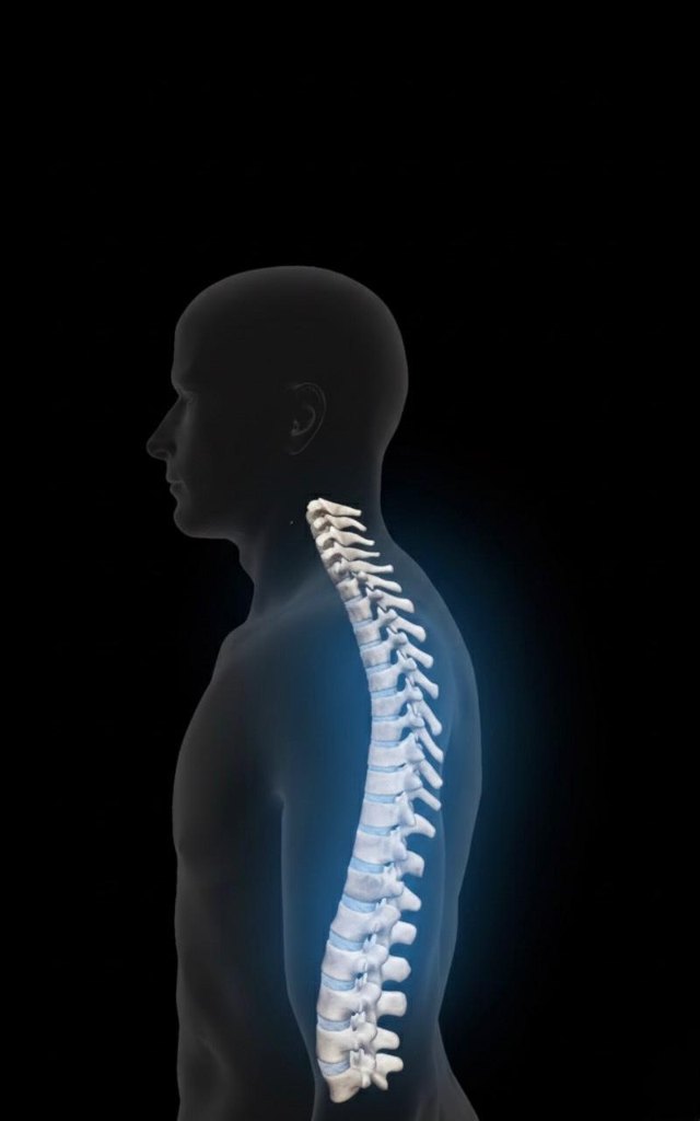
Spinal Cord Injury (SCI) causes permanent paralysis because the body cannot naturally regrow severed nerve fibers (axons) across the lesion site. With 302,000 Americans affected, fully restorative treatment remains elusive. University of Minnesota researchers are creating a revolutionary, custom-designed living bridge to restore function. They are combining advanced 3D printing with stem cell technology to produce functional segments of spinal cord tissue, offering unprecedented hope for reconnecting the nervous system.

Introduction

Imagine a wound that refuses to heal, not because of infection, but because the crucial connection across the gap is permanently severed. That is the devastating reality of a Spinal Cord Injury (SCI). Despite tremendous medical advancements, the problem of functional loss for the approximately 302,000 people affected by SCI in the U.S. remains a profound, heartbreaking challenge with no fully restorative treatment available.

The core issue? The body cannot naturally regrow the long, delicate nerve fibers (axons) across the lesion site, especially in chronic injuries, leading to permanent paralysis.

But what if we could build a custom-designed, living bridge? Researchers at the University of Minnesota have combined the power of advanced 3D printing with cutting-edge stem cell technology to do just that, creating tiny, functional segments of spinal cord tissue that offer an unprecedented pathway to restoring movement.

The Vision: Designing the 'Ideal Substrate'

For a long time, therapies focused on neuroprotection—trying to save the nerve cells immediately after the injury. This approach has proven largely ineffective for the long-term, chronic injuries that patients live with every day. A new strategy was needed: one that could establish a functional neural relay mechanism to bypass the damage entirely.

The researchers understood that simply injecting cells wouldn't work; the cells need structure and guidance. They needed an 'ideal scaffold', and they found it in Spinal Cord Organoids .

Why Organoids? Organoids are lab-grown, three-dimensional cellular assemblies that closely mimic the intricate anatomy of the host spinal cord. They are the most sophisticated, anatomically similar substrate that could be introduced into the injury site, providing the perfect environment for cells to grow and connect.

The Manufacturing Marvel: Precision 3D Bioprinting

To ensure the organoids could successfully bridge the gap and guide the nerve fibers in the correct direction (rostral and caudal, or up and down the spine), the team engineered a supporting structure using 3D bioprinting.

Building the Perfect Bridge:

  1. Structural Backbone: They chose silicone as the scaffold material. Why? Because of its exceptional biocompatibility (it won't be rejected by the body) and its non-degradable nature. This permanent framework provides the necessary structural support and mechanical stability for long-term study of the growing organoids.

  2. The Blueprint:  The resulting scaffold was miniature but highly complex: roughly 1.6 mm wide, 0.65 mm high, and 2 mm long. The key feature was the internal structure: three microscopic channels, each about 11.2 micro meter wide, which act as precision tunnels to direct nerve growth.

  3. The Living Component: The cell source was clinically relevant human iPSC-derived spinal neural progenitor cells (sNPCs). These are human stem cells, which can be derived from a patient’s own cells in future applications to potentially avoid immune rejection. These sNPCs, mixed in a specialized bioink (Matrigel-based), were precisely dispensed into the printed silicone channels using an extrusion-based multi-material printing system.

The printing process essentially created a tiny, pre-wired neural highway, where the physical structure guides the biological growth.

The Final Test: Locomotor Recovery and Connectivity

The ultimate goal was to prove that this complex, bio-printed structure could work in vivo (in a living organism).

  1. The Transplantation Protocol:

    1. The scaffolds were allowed to culture for 40 days in the lab until the sNPCs formed organized, functional organoids.

    2. "The finished structures (two assembled scaffolds) were then carefully implanted into the 1.8 mm gap (resulting from a severe complete transection) in a rat model's spinal cord. It is crucial to note that this is a model of severe injury, making successful recovery much more difficult than in typical hemisection models."

  2. The Evidence of Recovery: Stepping Forward:

    The data on functional recovery provides stark evidence for the effectiveness of the treatment. Recovery was quantified using the Basso, Beattie, and Bresnahan (BBB) Locomotor Score, the gold standard for assessing function in this model. The Organoid Group demonstrated a significant recovery, achieving a mean score of 8.4  0.93 at 12 weeks, which corresponds to the animals successfully taking occasional full weight-bearing steps. In contrast, the control groups showed minimal improvement: the Scaffold-Only Group reached only 3.6  1.25 (indicating minimal, mainly non-weight-bearing movement), and the Injury-Only Group showed extremely poor function with a mean score of 2.25  0.72. This difference highlights that the organoid-scaffold combination led to a robust, new functional pathway. While these scores are lower than those reported in less severe injury models, this achievement is remarkable given the severity of the complete transection model used.

  3. Proving the Connection: Motor Evoked Potentials

    1. To move beyond simple observation, the team measured the electrical signals moving through the spine using Motor Evoked Potentials (MEPs). This is the definitive proof of communication between the brain and the body below the injury.

      • The Organoid Group showed a Motor Evoked Potential (MEP) signal of 2.18  0.35 millivolts (mV), which is indicative of powerful, enhanced neural connectivity.

      • This signal was approximately double that of the scaffold-only group (1.07  0.17 mV) and nearly three times that of the injury-only group (0.83  0.18 mV).

This data confirms that the bio-printed bridge was not just a placeholder; it was an active communication relay.

  1. The Blueprint of Integration: Cell Fate

    When examining the implanted area, the researchers found the human cells (identified by the marker SC121+) had successfully integrated with the host rat tissue.17 After 12 weeks, the stem cells had matured:

    63.10%  2.31% of the cells differentiated into neurons, the essential signaling cells.

    20.21%  1.07% differentiated into oligodendrocytes, the cells that produce the insulating myelin sheath crucial for fast signal transmission.

Crucially, the neurons extended their nerve fibers in both the rostral and caudal directions from the scaffold, actively seeking out and connecting with the host's nervous system.18 This confirmed the physical creation of the functional neural relay, successfully linking the previously severed spinal segments.

Conclusion and the Horizon of Hope

This study from the University of Minnesota marks a monumental leap in regenerative medicine by achieving success through a novel, comprehensive strategy.

The novelty lies in the synergistic combination: marrying a multi-material 3D printing strategy with iPSC-derived regionally specific sNPCs to build functional organoid scaffolds.

The mechanism of recovery is clear: the scaffolds provided the essential structure and biological cues to guide the cells in forming a functional neural relay across a severe lesion gap, primarily through the integration of the right type of spinal interneurons.

For people with chronic SCI, the Clinical Outlook is one of renewed hope. This approach tackles the long-standing challenge of simultaneously providing both structure and substrate in one device. While the lack of current combination trials in the clinic highlights the work ahead, this research offers a clear and transformative path toward advancing therapies, suggesting that the era of irreversible paralysis may soon be coming to an end.

00
Engage with this post
Like, star, and comment to support the author and help others discover great content.
Loading...

More from this author

Microplastics:Our Arterial Threat
Microplastics cause heart harm. They trigger atherosclerosis, a buildup of arterial fats that impacts men and women differently. Recent genetic modeling confirms these tiny fragments move through our
GCF Backend for Zoho Campaigns
Integrating third-party services like Zoho Campaigns into a serverless backend is powerful, but it comes with unique challenges, especially around OAuth token management and specific API interaction q
Brain-Computer Interfaces: The Mind’s New Frontier
The boundary between human thought and machine intelligence is dissolving. Brain-computer interfaces (BCIs)—once the stuff of science fiction—are now a rapidly developing technology with potential to
A Smart Switch for Diabetes
A breakthrough protein called NNC2215 acts like “smart insulin”—auto-adjusting its activity based on blood sugar. Learn how it works, what the animal studies show, challenges ahead, and when it might
The Hope and Hype of Stem Cell Therapy
Revolutionary stem cell therapy promises to fix diseases by repairing or replacing tissues, offering a tangible alternative to conventional drug-based treatments.Stem cell-based therapy represents one